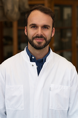
Dr. rer. nat. Jonas Keiler

Kontakt:
Universitätsmedizin Rostock AöR
Institut für Anatomie Rostock
Dr. rer. nat. Jonas Keiler
Gertrudenstraße 9
18057 Rostock
Tel.: +49 (0)381 - 494 8409
Raum: 3.23
E-Mail: jonas.keiler{bei}med.uni-rostock.de
Link zur Arbeitsgruppe: AG Keiler
Forschungsprojekt/Scientific profile
Entwicklung von nicht-kanzerogenen Gewebefixierungen für Histologie und Körperspendewesen
In vielen Bereichen der Biomedizin wird Formaldehyd als effizientes und kostengünstiges Fixierungsmittel zur Konservierung von biologischen Proben verwendet. Allerdings ist seine Anwendbarkeit durch seine bekannten krebserregenden und embryotoxischen Effekte stark eingeschränkt. Eine vielversprechende Alternative stellen hierbei nicht-toxische Konservierungsmittel aus der Lebensmitteltechnologie dar, mit denen wir in Vorversuchen bereits gute Ergebnisse hinsichtlich anti-mikrobieller Wirkung und mikroskopischen Gewebeerhalts erzielen konnten. Dank der Unterstützung der Deutschen Forschungsgemeinschaft (DFG) können wir unsere experimentellen Untersuchungen nun intensivieren.
Ex-vivo-Imaging in der Augenheilkunde
Die Mikrocomputertomografie (Mikro-CT) bietet ein weitgehend zerstörungsfreies Bildgebungsverfahren, das speziell zur Visualisierung und Analyse interner Strukturen des Ex-vivo-Auges eingesetzt wird. Im Gegensatz zu anderen hochauflösenden Bildgebungsverfahren ermöglicht die Mikro-CT eine dreidimensionale Erfassung komplexer Gewebestrukturen, wie der vorderen Augenkammer, in hoher Auflösung. Ziel des Projekts ist es, die Mikro-CT als Standardverfahren für die ophthalmologische Forschung zu etablieren und dadurch die Entwicklung neuer Therapien und Technologien zu unterstützen.
Morphologische und histologische Untersuchung humaner femoraler Venenklappen als Grundlage von zu entwickelnden Venenklappenimplantaten
Etwa fünf Prozent der über 50-Jährigen in Deutschland leiden an chronisch venöser Insuffizienz (CVI), die auf einer Mikrozirkulationsstörung der Gefäße infolge einer venösen Abflussbehinderung beruht. Im Rahmen dieses RESPONSE-Forschungsvorhabens wird an der Entwicklung eines innovativen Implantats zur Behandlung von insuffizienten Venenklappen gearbeitet. Die Anforderungen an ein perkutan, also minimalinvasiv zu applizierendes Venenklappenimplantat hinsichtlich geometrischer, mechanischer und morphologischer Parameter müssen präzisiert werden. Ziel ist daher die morphologische und histologische Charakterisierung von humanen Venenklappen u.a. mit röntgentomographischen und immunhistochemischen Methoden.
Topographische und immunhistochemische Charakterisierung von Sonden-Gefäßkontakten für die Optimierung von Schrittmacher- und Defibrillatorensonden
Die Sonden von Herzschrittmachern oder Defibrillatoren sind enormen dynamisch-mechanischen Langzeitbelastungen im venösen Blutstrom ausgesetzt. Deren polymere Isolationsmaterialien unterliegen zusätzlich Alterungsprozessen, die die Langzeitstabilität der Systeme verringern. Darüber hinaus erhöht die Fibrosierung und Gewebeadhäsion der Sonden die Extrahierbarkeit der Systeme im Fall von Defekten. Innerhalb dieses RESPONSE-Forschungsvorhabens werden deshalb neue Konzepte zur Optimierung der Sonden entwickelt. Ziel ist die topographische und immunhistochemische Charakterisierung der Adhäsionsstellen als Grundlage für die Entwicklung einer adhäsionsprotektiven Beschichtung, um Patienten zukünftig risikoarm mit langlebigen Implantaten versorgen zu können.
3D-Morphologie des menschlichen Eileiters
Bei etwa einem Viertel der Frauen mit unerfülltem Kinderwunsch liegt die Ursache im Verschluss des Eileiters, wobei der vordere Abschnitt am häufigsten betroffen. In-Vitro-Fertilisation ist zwar eine gängige Therapiemethode, wird aber von einigen betroffenen Paaren abgelehnt. Die Re-Kanalisierung des Eileiters mittels Katheter ist eine minimal-invasive Alternative, bei circa der Hälfte der Frauen tritt jedoch ein fibrotischer Wiederverschluss auf. Für eine Verbesserung der Behandlungsoptionen wird daher an der Universitätsmedizin Rostock ein Mikrostent entwickelt, der den Zweck hat, den Eileiter nach Re-Kanalisierung dauerhaft offen zu halten. Neben biologischen und biomechanischen Aspekten ist es dabei wichtig, auch geometrische Parameter für das Implantat festzulegen. Unser Ziel ist daher die Erfassung der Eileitermorphometrie auf der Basis von 3D-Röntgenmikroskopie und paraffinschnittbasierter Histologie.
Mitgliedschaften/Memberships
- German Zoological Society (Deutsche Zoologische Gesellschaft, DZG)
- Anatomische Gesellschaft
- Wikimedia Deutschland e.V.
- MikroMINT e.V.
- Verein zur Förderung der Auszubildenden und Studierenden an der Universitätsmedizin Rostock e.V.
Reviewing activities in scientific journals
- Histochemistry and Cell Biology
- Acta Amazonica
- Arthropod Structure & Development
- BMC Evolutionary Biology
- Journal of Morphology
- Zoology
- Zootaxa
- Marine Biology
- Journal of Crustacean Biology
- Annals of Anatomy
Publikationen
- Oster M, Reyer H, Hadlich F, Ponsuksili S, Wolf P, Wimmers K, Keiler J (2025). Parathyroid glands exhibit reduced parenchymatic chief cells and increased extracellular collagen as a response to a long-term low-phosphorus diet in pigs. Anim Nutr. 2025 Jul 10;22:471-482. doi: 10.1016/j.aninu.2025.04.007.
- Lebahn K, Keiler J, Schmidt W, Schubert J, Reumann M, Wree A, Grabow N, Kischkel S. Mechanical characterization of the human femoral vein wall and its valves by uniaxial tensile testing and hydrostatic compliance testing by optical coherence tomography (2025). Journal of the Mechanical Behavior of Biomedical Materials. https://doi.org/10.1016/j.jmbbm.2025.106938
- Keiler J, Bast A, Reimer J, Kipp M, Warnke P (2024). Quantitative and qualitative assessment of airborne microorganisms during gross anatomical class and the bacterial and fungal load on formalin-embalmed corpses. Scientific Reports 14, 19061 (2024). https://doi.org/10.1038/s41598-024-69659-y
- Antipova V, Heimes D, Seidel K, Schulz J, Schmitt O, Holzmann C, Rolfs A, Bidmon HJ, de San Román Martín EG, Huesgen PF, Amunts K, Keiler J, Hammer N, Witt M, Wree A (2024). Differently increased Volumes of Multiple Brain Areas in Npc1 Mutant Mice Following Various Drug Treatments. Frontier in Neuroanatomy. DOI: 10.3389/fnana.2024.1430790
- Steinhagen I, Brinker U, Kolbe V, Bingert R, Keiler J, Klussmann-Fricke B-J, Sokiranski R, Pirsig W, Begerock A-M (2024). The role of radiology in provenance research - experiences from the collaboration between radiology and anatomy at the University of Rostock and future perspectives. Fortschritte auf dem Gebiet der Röntgenstrahlen und bildgebenden Verfahren. DOI: 10.1055/a-2303-0312
- Keiler J, Stahnke T, Guthoff RF, Wree A, Runge J. Ex Vivo Micro-CT in Ophthalmology: Preparation and Contrasting for Non-invasive 3D-Visualisation. Klin Monbl Augenheilkd. 2023 Dec;240(12):1359-1368. English, German. doi: 10.1055/a-2111-8415 . Epub 2023 Dec 13. PMID: 38092003 .
- Runge J, Stahnke T, Guthoff RF, Wree A, Keiler J (2022): Micro-CT in ophthalmology: ex vivo preparation and contrasting methods for detailed 3D-visualization of eye anatomy with special emphasis on critical point drying. Quantitative Imaging in Medicine and Surgery 12(9):4361-4376. doi: 10.21037/qims-22-109
- Feifei L, Richter A, Runge J, Keiler J, Hermann A, Kipp M, Joost S (2022): Spontaneous hind limb paralysis due to acute precursor B cell leukemia in RAG1-deficient mice. Journal of Molecular Neuroscience. doi: 10.1007/s12031-022-02025-7
- Joost S, Schweiger F, Pfeiffer F, Ertl C, Keiler J, Frank M, Kipp M (2022): Cuprizone intoxication results in myelin vacuole formation. Frontiers in Cellular Neuroscience 16. doi: 10.3389/fncel.2022.709596
- Oster, M., Reyer, H., Keiler, J., Ball, E., Mulvenna, C., Ponsuksili, S., Wimmers, K. (2021): Comfrey (Symphytum spp.) as a feed supplement in pig nutrition contributes to regional resource cycles. Science of the Total Environment 796: 148988. Doi: 10.1016/j.scitotenv.2021.148988
- Oster, M., Reyer, H., Gerlinger, C., Trakooljul, N., Siengdee, P., Keiler, J., Ponsuksili, S., Wolf, P., Wimmers, K. (2021): mRNA profiles of porcine parathyroid glands following variable phosphorus supplies throughout fetal and postnatal life. MDPI Biomedicines 9 (454). doi: 10.3390/ biomedicines9050454
- Dierke, A., Borowski, F., Großmann, S., Brandt-Wunderlich, C., Matschegewski, C., Rosam, P., Pilz, N., Reister, P., Einenkel, R., Hinze, U., Keiler, J., Stiehm, M., Schümann, K., Bock, A., Chichkov, B., Grabow, N., Wree, A., Zygmunt, M., Schmitz, K-.P., Siewert, S. (2020): Development of a biodegradable microstent for minimally invasive treatment of Fallopian tube occlusions. Current Directions in Biomedical Engineering 6(3). doi: 10.1515/cdbme-2020-3019 .
- Rosam P., Stiehm, M., Borowski, F., Keiler, J., Wree, A., Öner, A., Schmitz, K.-P., Schmidt, W. (2020): Development of an in vitro measurement method for improved assessment of the side branch expansion capacity. Current Directions in Biomedical Engineering 6(3): 20203114. doi: 10.1515/cdbme-2020-3114.
- Keiler, J., Meinel, F., Ortak, J., Weber, M.-A., Wree, A., Streckenbach, F. (2020): Morphometric characterization of human coronary veins and subvenous epicardial adipose tissue – implications for cardiac resynchronization therapy devices. Frontiers in Cardiovascular Medicine 7: 611160. doi: 10.3389/fcvm.2020.611160.
- Keiler, J., Schulze, M., Dreger, R., Springer, A., Öner, A., Wree, A. (2020): Quantitative and qualitative assessment of adhesive thrombo-fibrotic lead encapsulations (TFLE) of pacemaker and ICD leads in arrhythmia patients - a post mortem study. Frontiers in Cardiovascular Medicine 7: 602179. doi: 10.3389/fcvm.2020.602179
- Oster, M., Reyer, H., Keiler, J., Ball, E., Mulvenna, C., Murani, E., Ponsuksili, S., Wimmers, K. (2020): Comfrey (Symphytum spp.) as an alternative field crop contributing to closed agricultural cycles in chicken feeding. Science of the Total Environment 742:140490. doi: 10.1016/j.scitotenv.2020.140490
- Kreft, D., Keiler, J., Grambow, E., Kischkel, S., Wree, A., Doblhammer, G. (2020): Prevalence and mortality of venous leg diseases of the deep veins. An observational cohort study based on German health claims data. Angiology 71(5):452-464. doi: 10.1177/0003319720905751
- Keiler, J.-A., Benecke, M., Keiler, J. (2020): Bone modifications by insects from the Early Pleistocene site of Untermassfeld. Kahlke, R.-D. (Ed.): The Pleistocene of Untermassfeld near Meiningen (Thüringen, Germany), Vol. 4: 1117-1132.
- Schubert, J., Schümann, K., Stiehm, M., Pfensig, S., Kischkel, S., Keiler, J., Wree, A., Schmidt, W., Schmitz, K.-P., Grabow, N. (2019): Numerical simulation of the functionality of a stent structure for venous valve prostheses. Current Directions in Biomedical Engineering 5(1):477-480. doi: 10.1515/cdbme-2019-0120
- Keiler, J., Seidel, R., Wree, A. (2019): The femoral vein diameter and its correlation with sex, age and BMI - a phlebological parameter with clinical relevance. Phlebology 34(1):58-69. doi: 10.1177/0268355518772746
- Pfensig, S., Kaule, S., Ott, R., Wüstenhagen, C., Stiehm, M., Keiler, J., Wree, A., Grabow, N., Schmitz, K.-P., Siewert, S. (2018): Numerical simulation of a transcatheter aortic heart valve under application-related loading. Current Directions in Biomedical Engineering 4(1): 185-189. doi: 10.1515/cdbme-2018-0046
- Stiehm, M., Kohse, S., Schümann, K., Kaule, S., Siewert, S., Oldenburg, J., Keiler, J., Grabow, N., Wree, A., Schmitz, K.-P. (2018): Hemodynamic influence of design parameters of novel venous valve prostheses. Current Directions in Biomedical Engineering 4(1):149-151. doi: 10.1515/cdbme-2018-0037
- Keiler, J., Schulze, M., Claassen, H., Wree, A. (2018): Human femoral vein diameter and topography of valves and tributaries: a post mortem analysis. Clinical Anatomy 31:1065-1076. doi: 10.1002/ca.23224
- Oster, M., Keiler, J., Schulze, M., Reyer, H., Wree, A., Wimmers, K. (2018). Fast and reliable dissection of porcine parathyroid glands - a protocol for molecular and histological analyses. Annals of Anatomy 219: 76-81. doi: 10.1016/j.aanat.2018.05.007
- Keiler, J., Schulze, M., Sombetzki, M., Heller, T., Tischer, N. Grabow, T., Wree, A., Bänsch, D. (2017): Neointimal fibrotic lead encapsulation – clinical challenges and demands for implantable cardiac electronic devices. Journal of Cardiology 70:7-17. doi: 10.1016/j.jjcc.2017.01.011
- Keiler, J., Wirkner, C.S., Richter, S. (2017): 100 years of carcinization – the evolution of the crab-like habitus in Anomura. Biological Journal of the Linnean Society 121:200–222. doi: 10.1093/biolinnean/blw031
- Wirkner, C.S., Göpel, T., Runge, J., Keiler, J., Klussmann-Fricke, B.-J., Huckstorf, K., Scholz, S., Miko, I., Yoder, M., Richter, S. (2017): The first organ based ontology for arthropods (Ontology of Arthropod Circulatory Systems – OarCS) and a semantic model for the formalization of morphological descriptions. Systematic Biology 66(5):754–768. doi: 10.1093/sysbio/syw108
- Keiler, J.; Richter, S., Wirkner, C.S. (2016): Revealing their innermost secrets: an evolutionary perspective on the disparity of the organ systems in anomuran crabs (Crustacea: Decapoda: Anomura). Contributions to Zoology 85(3):361-386. http://www.contributionstozoology.nl/vol85/nr04/a01
- Kaji, T., Keiler, J., Bourguignon, T., Miura, T. (2016): Functional transformation series and the evolutionary origin of novel forms: Evidence from a remarkable termite defensive organ. Evolution & Development 18(2):78-88. doi: 10.1111/ede.12179.
- Noever, C.; Keiler, J.; Glenner, H. (2016): First 3D reconstruction of the rhizocephalan root system using MicroCT. Journal of Sea Research 113:58-64. doi: 10.1016/j.seares.2015.08.002
- Jaszkowiak, K.; Keiler, J.; Wirkner, C.S.; Richter, S. (2015): The mouth apparatus of Lithodes maja (Crustacea: Decapoda) - form, function and biological role. Acta Zoologica 96:401-417. doi: 10.1111/azo.12135.
- Keiler, J.; Richter, S.; Wirkner C.S. (2015): The anatomy of the king crab Hapalogaster mertensii Brandt, 1850 (Paguroidea: Hapalogastridae) – new insights into the evolutionary transformation of hermit crabs into king crabs. Contributions to Zoology 84(2):149-165. http://www.contributionstozoology.nl/vol84/nr02/a04
- Keiler, J.; Richter, S.; Wirkner C.S. (2015): Evolutionary Morphology of the organ systems in squat lobsters and porcelain crabs (Crustacea: Decapoda: Anomala) – An insight into carcinization. Journal of Morphology 276:1-21. doi: 10.1002/jmor.20311
- Keiler, J.; Richter, S.; Wirkner C.S. (2013): Evolutionary Morphology of the Hemolymph Vascular System in Hermit and King Crabs (Crustacea: Decapoda: Anomala). Journal of Morphology 274:759–778. doi: 10.1002/jmor.20174
- Keiler, J.; Richter, S. (2011): Morphological diversity of setae on the grooming legs in Anomala (Decapoda: Reptantia) revealed by scanning electron microscopy. Zoologischer Anzeiger 250(4): 343-366. doi: 10.1016/j.jcz.2011.04.004
Curriculum vitae
| 2002-2008 | Studies of Biosciences at the University of Rostock, Germany |
| 08/2008 | Graduate degree (Dipl.-Biol.) at the Institute for Biosciences at the University of Rostock, Germany |
| 2008-2010 | Academic assistant (German Science Foundation project) at Institute for General and Advanced Zoology, University of Rostock, Germany |
| 2010-2013 | Academic assistant (German Science Foundation project) at Institute for General and Advanced Zoology, University of Rostock, Germany |
| 2008-2015 | Doctoral thesis at the University of Rostock, Germany (grade: 1,0 - magna cum laude) |
| 2015 | Academic assistant (RESPONSE research project) at Department of Anatomy, Rostock University Medical Center, Germany |
